Ever wonder why the outer ear is the way it is?
The ear consists of the outer, middle, and inner ears, making the entire organ much more extensive than we typically think. On top of that, an auditory nerve connects the ear to the brain, and a Eustachian tube links the middle ear to the top of the throat. The entire ear attaches to the temporal bone. Indeed, the anatomy of the outer ear is just the beginning of a more extensive construction.
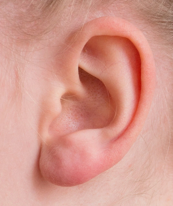
The Anatomy of the Outer Ear
Helix
The helix is the outermost rim of the ear that goes from the top to the lobe. It is the first “circuit” that channels sound waves closer to the eardrum.
Anti Helix
The anti helix is the protruding fold within the helix, particularly on the lower end of this large portion. On the upper end, you might notice two protruding stems that converge into the larger part. The upper stem is called the superior crus, and the lower is the inferior crus. The indentation between them is the triangular fossa.
Fossa
A fossa is any indentation in the ear. For example, the triangular fossa is such an indentation, as is the crease under the helix, which is called the scaphoid fossa.
Cymba Conchae
You might find yourself cleaning this part of the ear out sometimes, especially if you fidget. It is the pocket below the triangular fossa and above the ear hole, also known as the cavum concha. The helix wraps under the cymba conchae and ends next to the anti helix.
Cavum Conchae
You will find varying diagrams of the outer ear online. The cavum conchae, for instance, could also be called the concha cavum. In any case, this concha is the deep impression that leads to the ear canal.
Tragus
Two pieces of cartilage stick out on either side in front of the concha cavum. The one that is closer to the face is called the tragus. The other one is the anti tragus. The dip between them is called the intertragic notch.
Lobule
The lobule is what we typically call the ear lobe. It is the only part of the outer ear that does not contain cartilage. Some believe that it is there to help regulate the ear’s temperature.
The Function of the Outer Ear
All of these parts of the outer ear work together to funnel sound waves into the ear canal, also called the external acoustic meatus. The ear looks the way it does for a reason!
Trust the Experts at Clarity Audiology & Hearing Solutions
Need help with your hearing? Clarity Hearing can help. Clarity Audiology & Hearing Solutions is an independently owned and operated clinic that focuses on quality of care and personalized, friendly service to the surrounding areas of Ellicott City, Catonsville, Columbia. Our Doctors of Audiology are highly trained with advanced degrees and take the time to provide the personalized care and attention that you need and deserve. We provide advanced hearing aid options that are personally calibrated with cutting-edge digital technology to fit your hearing loss, your unique ear anatomy, and your individual listening needs.
You can call us today to schedule a FREE Hearing Protection Consultation at 410-698-6594! For more blog posts, news, and updates, follow us on Facebook, Twitter, Pinterest, and LinkedIn.